Imaging
Studies can include comprehensive imaging support provided in conjunction with local university laboratories. Our imaging capabilities are managed by veterinary radiologist and medical physicist support.
Transpharmation can provide MRI (1.5T, 3.0T, 7.0T, 9.4T) for rodents, cats, and dogs at all listed field strengths, CT (4-slice, 64-slice, and micro-CT), PET and micro-PET, SPECT, ultrasound, plain and contrast radiographs, QMR and DEXA.
Imaging Capabilities
-
Several imaging protocols can be used to quantify the efficacy of new therapeutics. These can be conventional anatomic measures of tumor size using MRI, ultrasound or radiographs. Other functional measures that can be used on a quantitative basis pre and post treatment as a measure of the therapeutic effect of a new agent include:
- FDG PET imaging of glucose metabolism in tumors. Reduced glucose metabolism is a measure of therapeutic success.
- Dynamic contrast enhanced MRI to quantify effect of agents on angiogenesis. Reduced angiogenesis is a measure of therapeutic success
-
- Dynamic contrast perfusion to assess blood brain barrier permeability and regional cerebral blood flow using either MRI or CT
- PET/CT imaging with additional coregistration to MRI images if required to quantify glucose uptake
- Imaging of brain tissues using standard PET radiopharmaceuticals eg F-18 DOPA
- Imaging of brain tissues with custom developed radiopharmaceuticals linked to specific naturally occurring brain chemicals or novel compounds
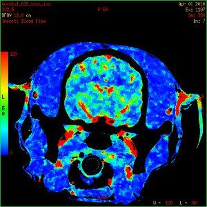
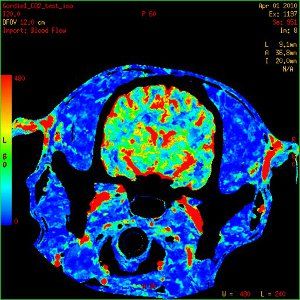
Dynamic perfusion CT images of a dog brain with cerebral blood flow (CBF) maps demonstrating an increase in CBF when end tidal CO2 is increased from 35mmHg (A) to 68mm Hg(B).
-
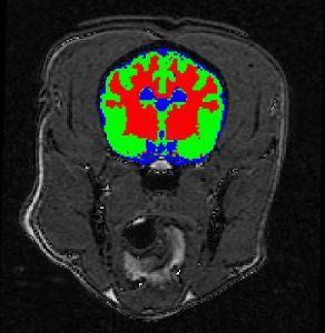
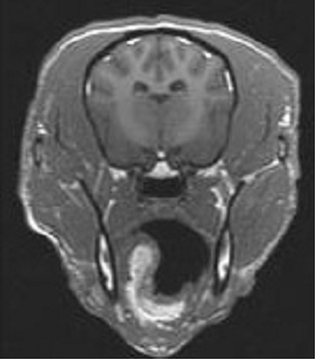
Dog brain segmented into grey matter, white matter and cerebrospinal fluid Transpharmation has experience in image processing of all modalities. Image processing extracts quantitative data from images. This can range from basic morphometric measurements made from images such as bone length or hippocampal volume to complex segmentation based on image signal intensities.
Project specific image processing routines are developed using scripts in standard software packages or can be custom developed according to the needs of the project. Interpretation of the data is routinely provided.
We are able to co-register images to enable low resolution quantitative images to be co-located with high resolution anatomical mages to allow accurate regions of interest to be drawn of the low resolution images. Brain images can be segmented into grey matter, white matter and cerebrospinal fluid. Regional blood flow and blood volume can be determined using either an MRI or a CT acquisition. Time activity curves can be generated from nuclear medicine studies to determine first pass kinetics.
-
Interventional imaging provides minimally invasive methods of collecting tissue or depositing cells in virtually any organ system. We have extensive experience in interventional procedures with all imaging modalities. The complexity can range from simple collection of fluid or cells to implantation of cells within a fetus to create chimera or place a device within an organ or vessel using an endovascular approach. Biopsies can be collected from both bone and soft tissues. We can also develop instrumentation and methodology for unique procedures.
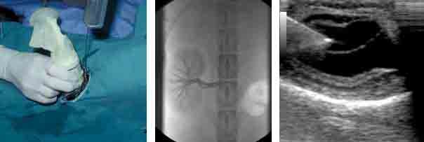
From left to right: Dog chest radiograph; image of dog prostate with benign prostatic hypertrophy; dog kidney with chronic infection.
-
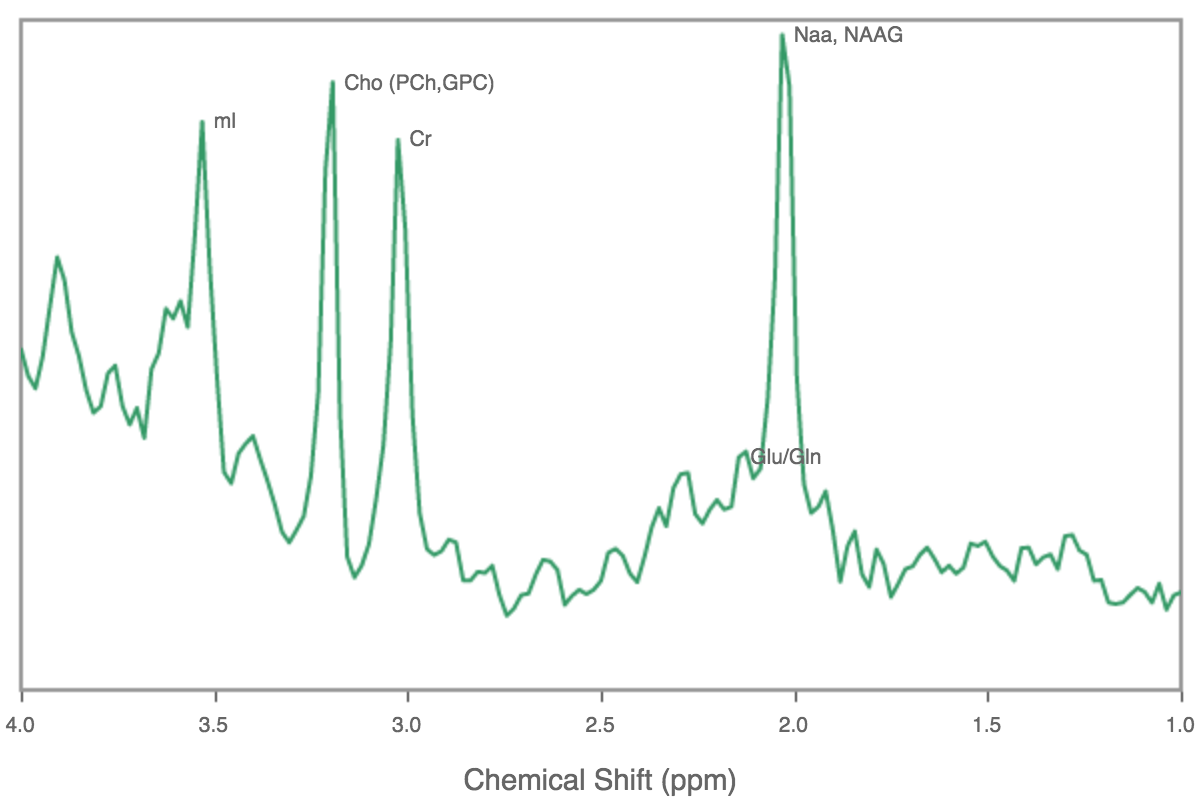
Quantification of changes in brain chemistry associated with the development and decline of neural function in response to therapeutics 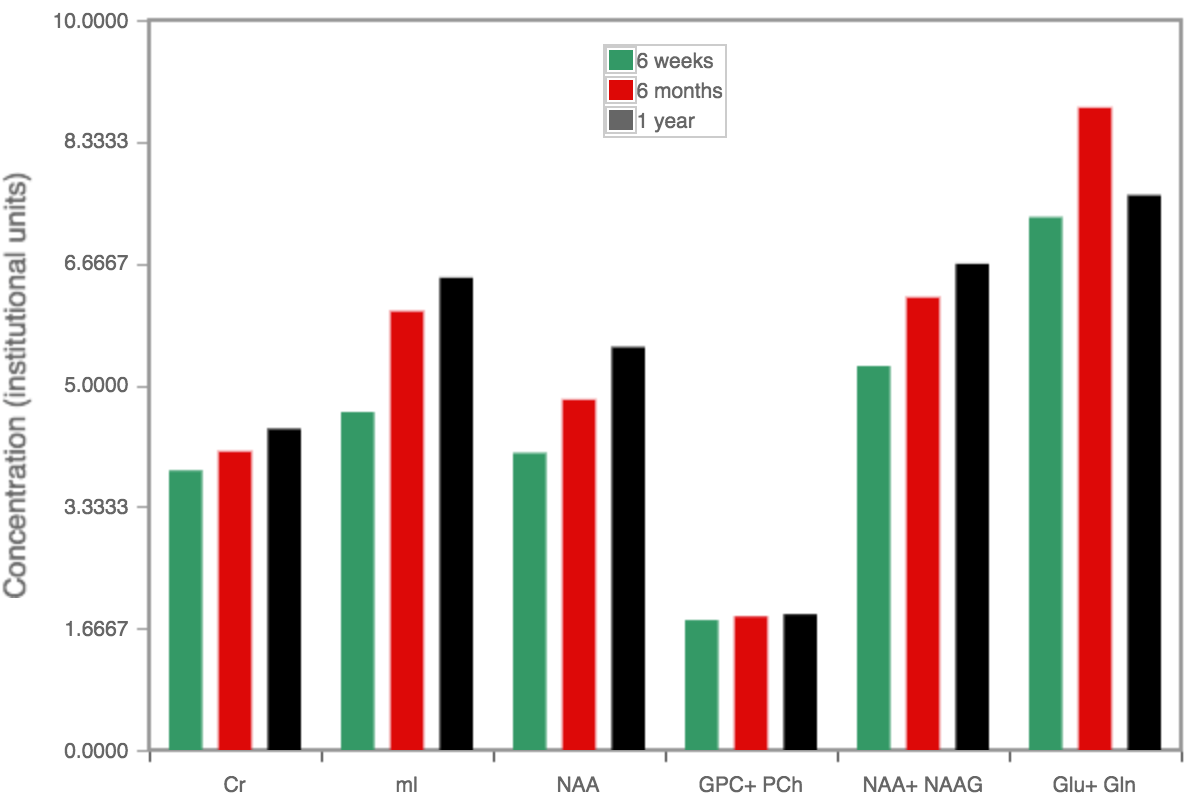
Magnetic resonance spectroscopy data showing changes in brain metabolites associated with brain development in growing beagles, including markers of neuronal health. Effects of therapeutics can be assessed in attenuating or improving brain development 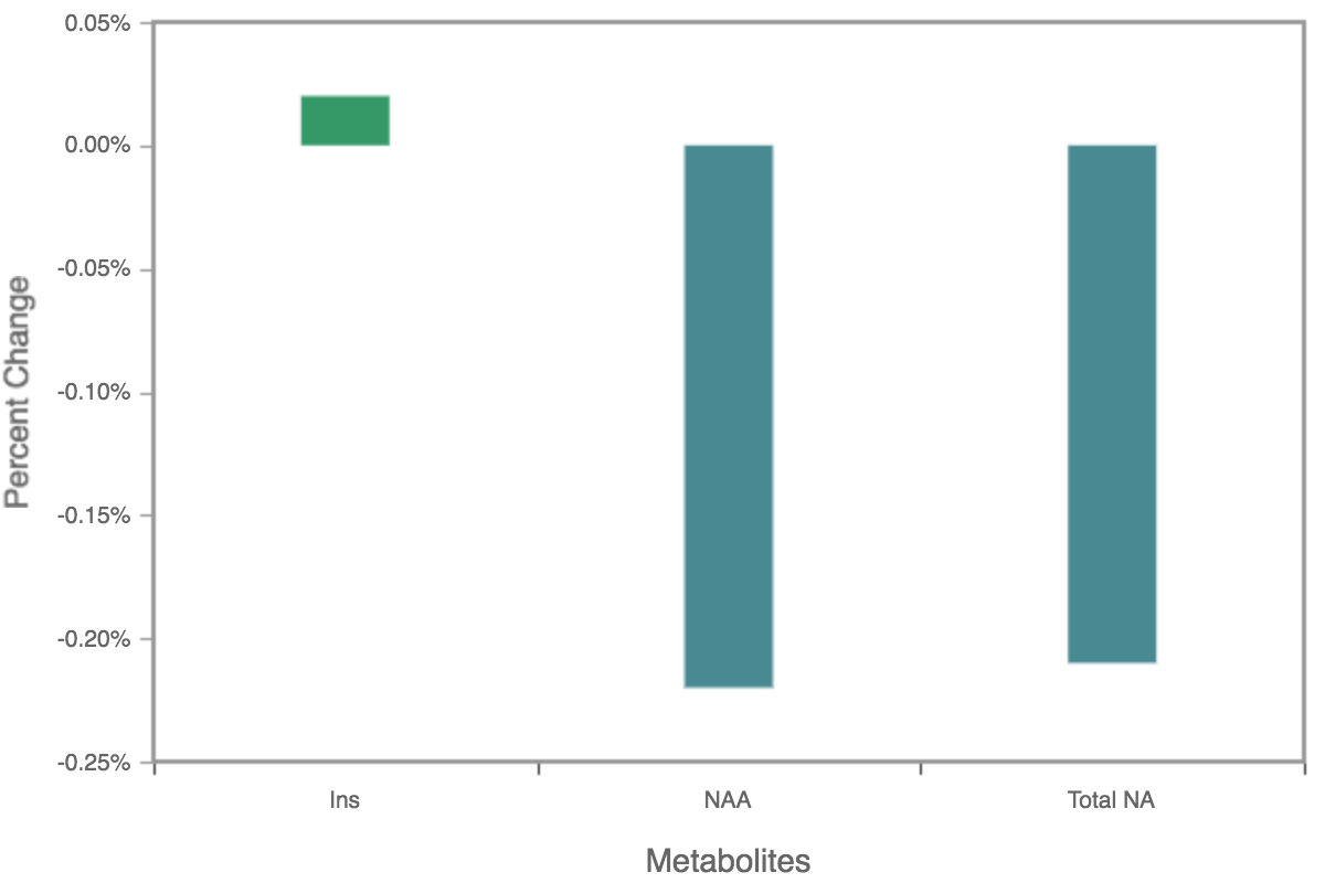
Data showing change in concentration of brain metabolites in 10 year old compared to 2 year old beagle dogs -
Depending on the research question to be answered several different imaging modalities can be used in musculoskeletal imaging. Magnetic Resonance Imaging provides images of the greatest range of tissues including bone marrow and changes in the internal structure of a joint, including articular cartilage changes, whereas CT can provide detailed images of bone structure.
Our range of magnets available allows MR imaging of beagles at up to 9.4T. Plain radiographs can illustrate basic morphometric changes with either contrast radiography or ultrasound adding details of the internal structure of joints. Nuclear medicine studies provide quantitative information on bone metabolic turnover and studies can be focused on a specific area or can include the entire skeletal system.
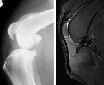
From left to right: Radiograph of a dog knee with osteoarthritis; MR image of a normal dog knee.
-
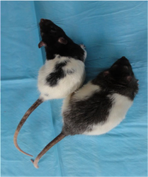
Quantitative magnetic resonance (QMR) is a non invasive method of determining body composition in awake animals. The principles of the method are similar to magnetic resonance imaging (MRI), except that the output is numerical rather than an image. Non sedated rodents are restrained in a Plexiglas tube that is placed within the bore of a low field magnet. Data collection takes two minutes, allowing a high throughput and repeated measurement for longitudinal study designs. During the data collection, hydrogen atoms in fat and lean tissue respond differently to radiofrequency energy, allowing the determination of the subject’s fat, lean and total body water mass.
-
Our comprehensive imaging capabilities allow evaluation of any organ or body system. In addition to the sophisticated cross sectional imaging modalities we have extensive experience in plain and contrast radiography and ultrasound, including endocavitory, evaluation. These modalities continue to have applications in the development of new therapeutic applications and we can help you to select the most appropriate and cost effective imaging method for your study. Morphometric measures can be made from the images and some measures of echogencitiy can be made from ultrasound images.
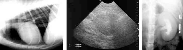
From left to right: Dog chest radiograph; image of dog prostate with benign prostatic hypertrophy; dog kidney with chronic infection.
-
- MRI or CT to demonstrate structural changes in response to interventions
- T1, T2. PD sequences
- Diffusion Tensor Imaging to evaluate changes in myelination
- Quantitative and qualitative assessment of morphological features or regions of interest by image processing
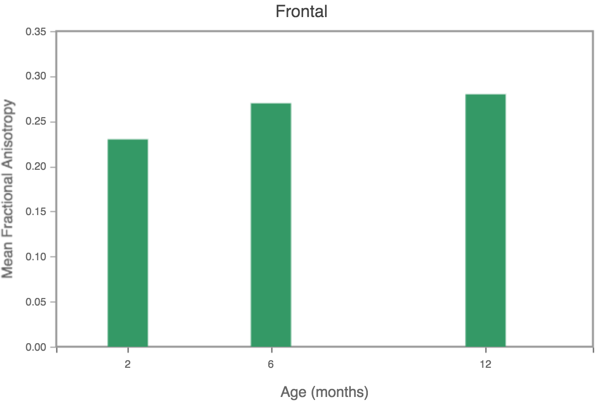
Diffusion Tensor Imaging data showing increasing fractional anisotropy in the frontal lobe, implying increasing myelination with increasing age in young beagle dogs. 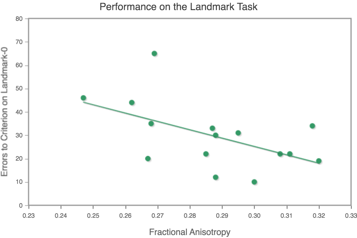
Data indicating performance on the landmark allocentric learning improves with increased myelination in one year old beagle dogs. -
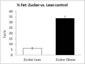
Whole body imaging protocols are used for assessing the effects of therapeutics on metabolism and feeding behavior. Using dual energy x-ray absorptiometry (DEXA) body composition can be determined non invasively in longitudinal studies. These studies give body composition in terms of the percentage of fat, lean and bone tissue. Body composition is an important component in obesity studies.
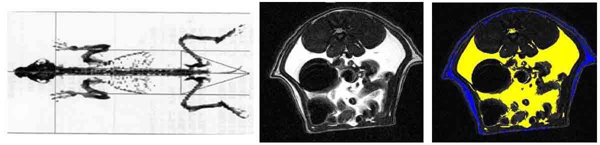
From left to right: DEXA image of a dog; MR Image of rat abdomen; MR image segmented into visceral (yellow) and subcutaneous (blue) fat.